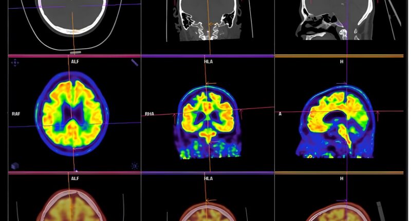
A Harvard Medical School-led research team has developed an AI tool that distinguishes between two look-alike cancers found in the brain with more than 98 per cent accuracy during surgery.
The tool, called PICTURE (Pathology Image Characterisation Tool with Uncertainty-aware Rapid Evaluations), tells apart glioblastoma, the most common and aggressive brain tumour, from primary central nervous system lymphoma (PCNSL), a rarer cancer often mistaken for glioblastoma. While both can appear in the brain, glioblastoma arises from brain cells, whereas PCNSL develops from immune cells.
Correctly identifying these look-alike tumours during surgery can expedite critical treatment choices. Glioblastoma requires surgical removal of cancerous tissue, while PCNSL should be left behind and treated with radiation and chemotherapy instead. Inaccurate or delayed diagnosis can lead to unnecessary surgery and delays in proper treatment.
“Our model can minimise errors in diagnosis by distinguishing between tumours with overlapping features and help clinicians determine the best course of treatment based on a tumour’s true identity,” said study senior author Kun-Hsing Yu, associate professor of biomedical informatics in the Blavatnik Institute at HMS and HMS assistant professor of pathology at Brigham and Women’s Hospital.
The model was evaluated on 2,141 brain pathology slides collected worldwide and tested across five international hospitals in four countries. In every case, the AI model outperformed existing AI tools and traditional frozen-section assessment, the standard of care for real-time tumour typing.
The tool includes an uncertainty detector that signals when it is unsure in its judgement and flags tumours the model has not encountered before for human review. PICTURE identified samples belonging to 67 CNS cancers that were neither gliomas nor lymphomas.
PICTURE outperformed human pathologists in hard-to-distinguish tumours. In tests, human specialists showed significant disagreement on difficult diagnoses, with some tumour types misdiagnosed 38 per cent of the time. PICTURE correctly identified all these cases.
The work, supported in part by the National Institutes of Health, is described in Nature Communications on 29 September. The AI model is publicly available for other scientists to use and build upon. Each year, more than 300,000 people worldwide are diagnosed with tumours in the brain or central nervous system, and more than 200,000 deaths occur as a result.


