
The brainstem drives the body’s most essential functions — consciousness, breathing, heart rate and sleep — yet for decades, medical imaging has struggled to see it clearly.
Now, a team of researchers from MIT, Harvard and Massachusetts General Hospital has unveiled an AI-powered tool capable of mapping this “unexplored” region with unprecedented clarity.
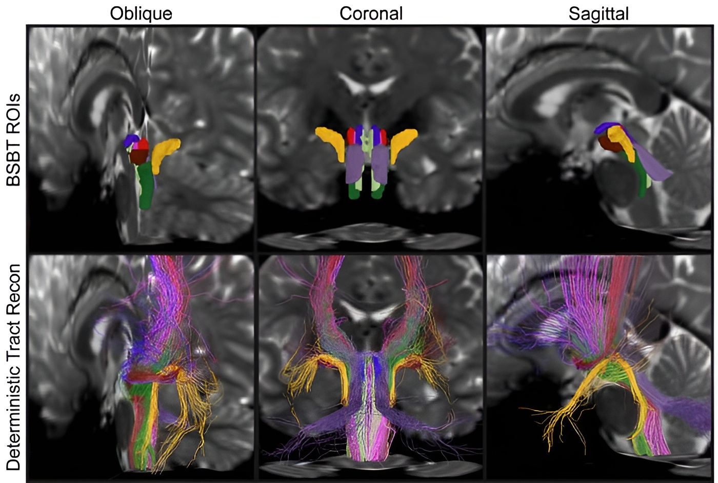
In a study published in the Proceedings of the National Academy of Sciences, the team describes how their new software, the BrainStem Bundle Tool (BSBT), can resolve eight distinct nerve bundles that were previously invisible to standard MRI scanners.
“The brainstem is a region of the brain that is essentially not explored because it is tough to image,” says Mark Olchanyi, a doctoral candidate at MIT and the study’s lead author. “People don’t really understand its makeup from an imaging perspective.”
The motion problem
The challenge has always been one of movement and scale. The brainstem is small, complex, and constantly jiggled by the pulsing of blood and the rhythm of breathing, blurring the “white matter” cables that connect the brain to the body.
To overcome this, Olchanyi trained a “convolutional neural network” on high-resolution scans, teaching the AI to ignore the noise and identify the specific pathways of the nerve bundles.
“The brainstem is one of the body’s most important control centres,” says Emery N. Brown, a professor at MIT and co-senior author. “Mark’s algorithms… offer us new access to vital physiological functions such as control of the respiratory and cardiovascular systems.”
The potential for the tool was highlighted by the case of a 29-year-old man who spent seven months in a coma following a traumatic brain injury.
By applying the AI tool to scans taken throughout his recovery, doctors could see that his brainstem bundles had been displaced but not severed. As the man recovered, the software tracked the lesions shrinking by a factor of three and showed the nerve bundles physically moving back into their correct positions.
Diagnosing disease
Beyond trauma, the tool showed promise in diagnosing neurodegenerative conditions. When applied to scans of patients with Parkinson’s disease, multiple sclerosis (MS) and Alzheimer’s, the AI detected distinct patterns of damage.
For example, while Alzheimer’s patients showed decline in only one bundle, those with Parkinson’s showed degradation in three. In MS patients, the tool identified significant volume loss in specific pathways.
The researchers believe the tool could become a “key adjunct” for doctors, offering a non-invasive way to assess brainstem health and predict recovery.