
A new AI-assisted brain atlas that can help visualise the human brain in unprecedented detail has been developed by UCL researchers, enabling the analysis of MRI scans of living patients in a matter of minutes at a level of detail not previously possible.
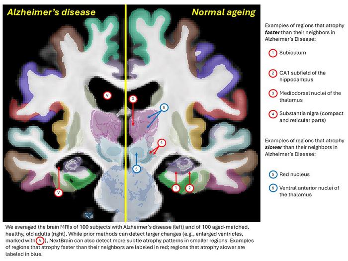
The human brain comprises hundreds of interconnected regions that drive thoughts, emotions and behaviours. Existing brain atlases can identify major structures in MRI scans, but their finer sub-regions remain hard to detect. These distinctions are important because sub-regions are affected differently during the progression of Alzheimer’s disease. The findings were published in Nature.
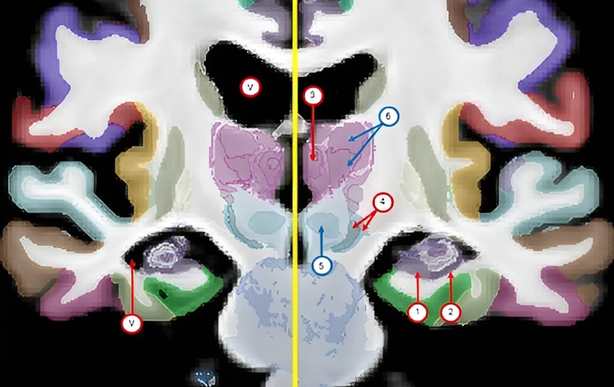
The atlas, called NextBrain, took the research team six years to build through a painstaking process using post-mortem tissue from five human brains. Each brain was dissected and sectioned into 10,000 pieces, stained to help identify brain structures, photographed under a microscope, and then reassembled into a 3D digital model.
AI was used to help align the microscope images and the MRI scans, accounting for the differences between the two techniques and ensuring that the pieces did not overlap or have gaps in between them. A total of 333 brain regions were then labelled on the digital 3D models of each of the five brains, a process greatly accelerated by AI that would have taken decades if done manually.
“NextBrain is the culmination of years of effort to bridge the gap between microscope imaging and MRI. By combining high-resolution tissue data with advanced AI techniques, we’ve created a tool that allows researchers to analyse brain scans in a level of detail that was previously unattainable,” said Dr Juan Eugenio Iglesias, senior author of the study from UCL Medical Physics & Biomedical Engineering and Massachusetts General Hospital/Harvard Medical School.
The resulting atlas, which is an average of the five brain models, is generalisable to all adult humans, meaning it can be used to automatically infer details from MRI scans of living or deceased subjects.
NextBrain was successfully tested on thousands of MRI datasets, demonstrating the ability to reliably identify brain regions across diverse imaging conditions and scanner types. In one experiment, the researchers applied NextBrain to over 3,000 MRI scans of living individuals to investigate age-related changes in brain volume.
“Our goal by building this atlas was to enable researchers to identify hundreds of brain regions in living patients quickly and consistently, while maintaining the fine-grained anatomical accuracy of microscope data,” said Dr Zane Jaunmuktane, an author of the study from UCL Queen Square Institute of Neurology and the Queen Square Brain Bank for Neurological Disorders.
The creators of the atlas, which is freely available, hope it will ultimately help to accelerate discovery in brain science and its translation into better diagnosis and treatment of conditions such as Alzheimer’s. All underlying data, tools and annotations used in NextBrain have been released openly through the FreeSurfer neuroimaging platform.